2024-12-22T13:49:13  read more
Dr Gautam Sonography Center and Clinic 2024-12-22T13:49:13 2024-12-22T13:49:13
Dr Gautam Sonography Center and Clinic 
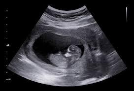
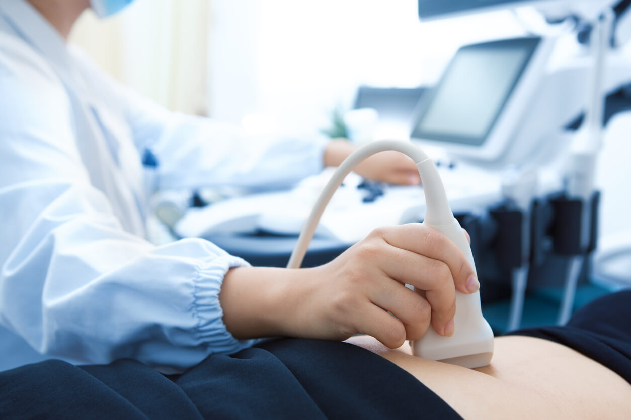
How Sonography Helps in Monitoring Pregnancy: A Guide for Expecting Parents Pregnancy is an exciting and sometimes uncertain journey for expecting parents. With so many questions about the baby's health, growth, and development, regular monitoring becomes essential to ensure everything is progressing smoothly. Sonography (or ultrasound ) plays a crucial role in providing accurate information and peace of mind during pregnancy. It’s a non-invasive, safe, and highly effective way to monitor both the mother’s and baby’s health, offering invaluable insights at various stages of pregnancy. In this guide, we’ll explore how sonography helps in monitoring pregnancy , the different types of ultrasounds used during pregnancy, and why they’re so important for expecting parents. What is Sonography and How Does it Work During Pregnancy? Sonography uses high-frequency sound waves to create images of the inside of the body. During pregnancy, ultrasound is used to visualize the baby, monitor its development, and check the health of both the baby and the mother. The process is non-invasive, meaning it doesn’t involve any surgery or needles, and it’s entirely safe for both the mother and child. A gel is applied to the mother’s abdomen, and a small transducer (a handheld device) is moved over the belly. The sound waves emitted by the transducer bounce off the tissues and organs, which are then converted into images on a screen. These images provide crucial information for doctors and expecting parents. Why is Sonography Important During Pregnancy? Confirming the Pregnancy The first ultrasound, often done in the first trimester , is essential to confirm the pregnancy and rule out any potential complications, such as ectopic pregnancy (when the fetus implants outside the uterus). This early ultrasound can also determine the number of fetuses (single or multiple pregnancies) and help estimate the due date. Monitoring Fetal Development and Growth As the pregnancy progresses, sonography is used to track the baby’s growth and development . Ultrasound scans provide clear images of the baby’s size , weight , heartbeat , and position in the uterus. These scans can help detect any growth issues, such as intrauterine growth restriction (IUGR) , ensuring the baby is developing at the right pace. Evaluating the Placenta Sonography also plays a key role in assessing the health of the placenta , which is vital for the baby’s nutrition and oxygen supply. Issues such as placental abruption (separation from the uterine wall) or placenta previa (when the placenta covers the cervix) can be identified through ultrasound, allowing the doctor to monitor and manage these conditions. Checking the Baby's Heartbeat and Movement Ultrasound allows doctors to monitor the fetal heartbeat and movement , which are strong indicators of the baby’s well-being. A healthy heartbeat (typically between 110-160 beats per minute) is one of the first signs that the baby is thriving. Fetal movement is another positive sign that indicates good health, and ultrasound helps doctors track this vital aspect of fetal development. Detecting Birth Defects and Abnormalities Ultrasound scans can detect birth defects and genetic conditions . The anomaly scan performed around 18-20 weeks of pregnancy checks for common conditions like Down syndrome , spina bifida , and cleft lip . This scan also helps assess the development of major organs such as the brain , heart , kidneys , and spine , allowing for early detection of any potential problems. Assessing Amniotic Fluid Levels The amount of amniotic fluid surrounding the baby is critical for its development. Ultrasound can measure the fluid levels and ensure they’re within normal ranges. Low amniotic fluid can indicate problems such as placental insufficiency or fetal dehydration , while too much fluid can be a sign of gestational diabetes or fetal abnormalities . Monitoring these levels through sonography helps prevent complications and allows for timely interventions. Checking the Cervix and Uterus Sonography also helps assess the cervix and uterus , checking for signs of premature dilation or other issues. Cervical insufficiency (when the cervix opens too early) can lead to preterm labor, so it’s important to monitor its length and condition through ultrasound. Position of the Baby Later in pregnancy, ultrasound helps doctors monitor the position of the baby, ensuring it’s in the proper position for delivery. Most babies are in the head-down position by the time of delivery, but some may be in a breech position (feet or buttocks first), which may require a caesarean section . Early detection allows expecting parents to explore options for turning the baby if needed. Types of Ultrasound Scans During Pregnancy Early Pregnancy Ultrasound This ultrasound is typically done around 6-8 weeks to confirm the pregnancy and check for an ectopic pregnancy. It can also provide an early estimate of the due date. Dating Ultrasound Usually performed between 8-14 weeks, this ultrasound helps establish an accurate due date by measuring the baby’s crown-to-rump length . First Trimester Screening Performed between 11-14 weeks, this ultrasound helps screen for potential chromosomal abnormalities like Down syndrome and provides a detailed assessment of the baby’s development. Anomaly Scan This ultrasound is performed around 18-20 weeks to check for birth defects or genetic conditions and assess the baby’s organs and growth . Growth Scan Done in the later stages of pregnancy, this ultrasound monitors the baby’s growth , position , and amniotic fluid levels to ensure everything is progressing normally. Biophysical Profile (BPP) This test is done in the third trimester to assess the baby’s movement , muscle tone , breathing , and the amount of amniotic fluid , helping doctors evaluate the baby’s well-being. Benefits of Sonography for Expecting Parents Peace of Mind : Regular ultrasounds reassure parents about the health and development of their baby. They can see their baby’s heartbeat, movement, and development, which is a source of comfort. Early Problem Detection : Sonography helps detect potential issues early, which can lead to better management and treatment. Early detection of complications like growth restriction or placental issues can be life-saving. Bonding with Your Baby : Ultrasound scans give expecting parents a chance to see their baby for the first time, creating an emotional connection and anticipation for the birth. Conclusion: The Vital Role of Sonography in Pregnancy Sonography is a powerful tool in monitoring pregnancy , offering crucial insights into both the health of the mother and the baby . Regular ultrasound scans allow for early detection of potential problems, ensuring the best possible outcomes for both mother and child. From early pregnancy confirmation to fetal health monitoring in the third trimester, ultrasound plays a vital role in every stage of pregnancy. At Dr. OP Gautam Sonography Center , we offer advanced ultrasound services to ensure the health and well-being of both the mother and the baby. With experienced sonologists and the latest ultrasound technology, we’re dedicated to providing accurate, reliable, and compassionate care for expecting parents. Book your ultrasound appointment today and take the first step toward a healthy pregnancy!